If I had to pick the single most over-requested scan in my career, it would be the MRI Lumbar Spine.
Lower back pain is extremely common. Most cases are due to muscle strain or posture and improve with rest and simple care. But when pain persists, spreads to the leg, or is accompanied by warning signs, doctors may suggest a spine MRI.
Here's what I tell every patient: don't rush into a spine MRI for simple back pain. Let me explain when it's truly useful, when it's not, and what it can show.
When You Do NOT Need a Spine MRI (Yet)
For simple, recent back pain without red flags, I usually advise against immediate imaging. Here's why:
- Pain for less than 4–6 weeks
- No leg pain, numbness, or weakness
- No bladder or bowel problems
- No fever, weight loss, or cancer history
In these situations, I recommend:
- Rest, gentle movement, and physical therapy
- Pain relief and anti-inflammatory medicines
- Posture and ergonomics changes
Imaging (including spine MRI) is typically delayed because:
- Many people have disc changes on MRI without symptoms—I see this all the time. A disc bulge doesn't always mean it's causing your pain.
- Early MRI can lead to unnecessary treatment—surgery for something that might have resolved on its own.
- Most acute back pain improves on its own—80-90% of patients get better within 6 weeks with conservative care.
I've seen patients who insisted on MRI after one week of back pain, found a small disc bulge (which is normal for their age), and then worried unnecessarily. Sometimes, waiting is the right medical decision.
When a Spine MRI IS Recommended
Red Flags
These are the signs that make me say "yes, we need imaging now":
- Numbness or weakness in the leg or foot—this suggests nerve compression
- Sciatica: pain radiating down the leg (often below the knee)—classic sign of disc pressing on nerve
- Bladder or bowel control problems (retention, incontinence)—this is a medical emergency
- Saddle numbness (perineal area)—another emergency sign
- Progressive weakness or foot drop—nerve damage is happening
- Fever with back pain (possible infection)—we need to rule this out
- History of cancer with new back pain—could be metastasis
- Significant trauma—fracture needs to be ruled out
- Pain that doesn't improve after 6 weeks of treatment—time to look deeper
If you have any of these, don't wait. Come to our centre in Thrissur or Ernakulam, and we'll prioritize your scan.
Other Indications
- Planning spine surgery or injections—surgeons need detailed images
- Pre-employment or insurance evaluation—sometimes required
- Monitoring known disc or stenosis—follow-up scans
- Persistent pain despite conservative treatment—after we've tried everything else
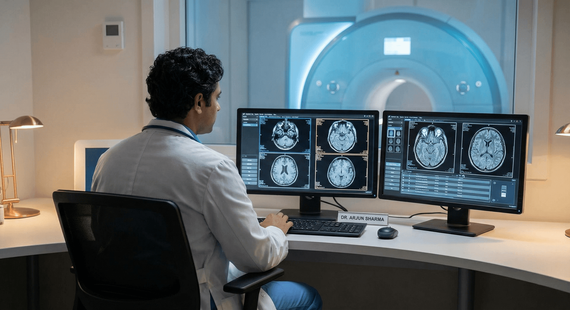
What Can a Spine MRI Detect?
When I review spine MRI scans, here's what I'm looking for:
Disc Bulge
Disc protrudes slightly beyond its normal space. Common; may or may not cause symptoms. I see this in most patients over 40—it's often just aging, not pathology.
Herniated Disc (Slip Disc)
Disc material pushes out and can press on nerves. Can cause sciatica, leg pain, numbness, weakness. This is when symptoms match the scan findings—that's when treatment becomes necessary.
Spinal Stenosis
Narrowing of the spinal canal. Often from arthritis; can cause leg pain when walking. Common in elderly patients—I see this frequently.
Spondylitis / Spondylosis
Degenerative changes: bone spurs, disc wear. Common with age; severity varies. This is normal aging, but sometimes it causes symptoms.
Other Conditions
- Infection (discitis, epidural abscess)—rare but serious
- Tumors (primary or metastatic)—uncommon but important to catch
- Fractures (e.g., vertebral compression)—especially in osteoporosis
Disc Bulge vs Herniation: What's the Difference?
A disc bulge is a generalized protrusion that's often age-related and may or may not cause symptoms. Herniation refers to a focal protrusion with a higher chance of nerve compression. When disc material breaks through the outer layer, it's called an extrusion. Sequestration occurs when a disc fragment becomes free in the spinal canal—this is the most severe form and often requires surgical attention.
The report describes size, location, and relationship to nerves. Your doctor uses this to plan treatment. I always explain this to patients—not every disc problem needs surgery. Many improve with physiotherapy and time.
What to Expect During a Spine MRI
When you come to our centre:
- Duration: About 20–40 minutes for lumbar spine
- Position: Lying on your back
- Stillness: Important for clear images—this is the hardest part
- Noise: Loud knocking; ear protection is provided
- Comfort: Wide-bore MRI at Magnus helps reduce claustrophobia; a relative can stay with you
If you have severe back pain, lying still can be challenging. Let us know—we can adjust positioning or discuss pain management options.
Summary
Spine MRI is valuable when lower back pain lasts, spreads to the leg, or comes with numbness, weakness, or bladder/bowel changes. For simple strain without red flags, conservative treatment first is often best. When needed, spine MRI clearly shows disc bulge, herniation, stenosis, and other causes of nerve compression.
Magnus Diagnostics offers spine MRI at Irinjalakuda and North Paravur with AI-enabled 1.5T scanners.
If you have persistent back pain with red flags, don't delay. Call +91 89031 01010 or visit our MRI services page to schedule your scan. But if it's simple back pain that just started, try rest and physiotherapy first—you might save yourself time and money.